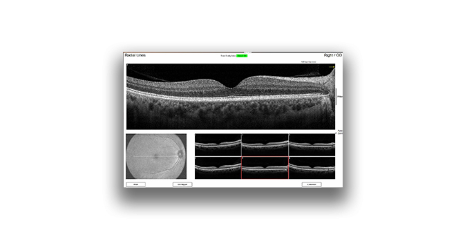
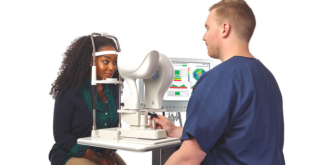
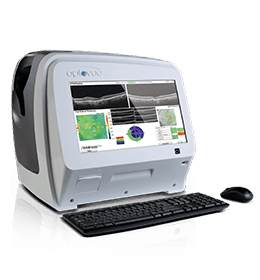
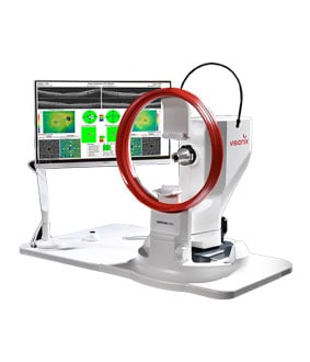
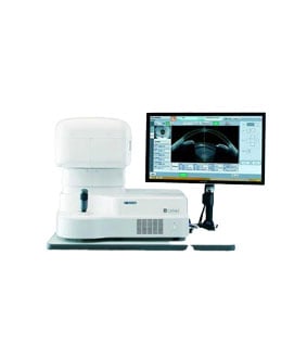
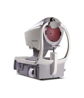
The iVue80 OCT is the newest innovation from the creators of OCT technology. At 80,000 A-scans per second, it is 3x faster than the original iVue for improved efficiency and enhanced image quality. Scan acquisition has been simplified with real-time en-face imaging displaying a 12x9 mm view of the retina during acquisition to assist the operator in scanning the desired location and new reports with a wider field of view for enhanced capabilities.
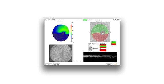
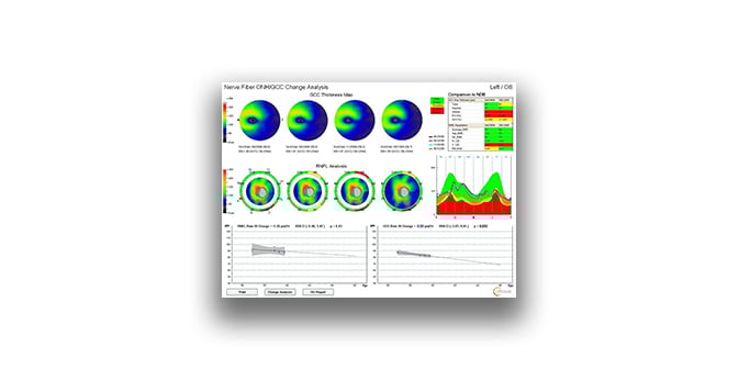
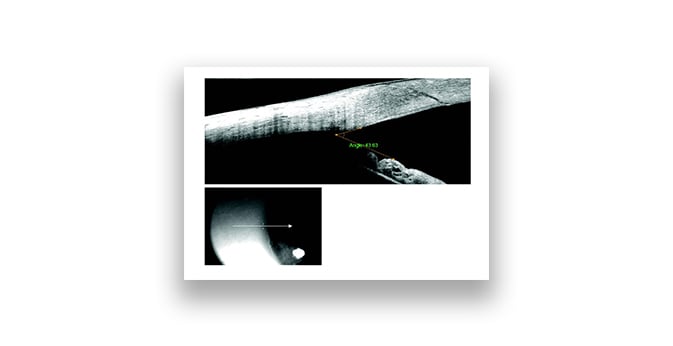
It is a powerful clinical tool that transforms the way you assess the retina, optic disc and cornea. Quantify the thickness of the retina, nerve fiber layer, ganglion cell complex (GCC) and the cornea. Track change and predict trends in RNFL and GCC thickness and precisely measure angles to aid in disease diagnosis.
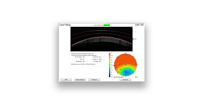
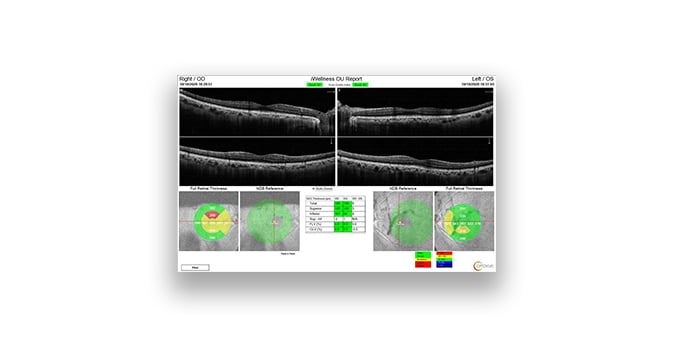
The iVue80 system delivers many Optovue exclusives such as ganglion cell complex (GCC) analysis with focal loss volume (FLV%) and global loss volume (GLV%) metrics, the iWellnessExam® and Vault Mapping for specialty lens fitting.
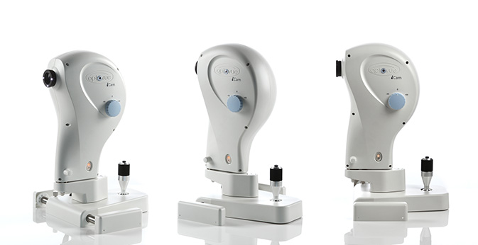
View Related Products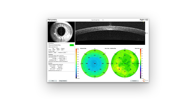

.png)
.png)
.png)


/S260%20S260S%20S280S%20(1).png)
.png)
%20(1).png)


.png)


.png)